University of Colorado School of Medicine
Dahl Lab
Using chromatin dynamics to better understand and treat pediatric brain cancers
About the lab
We study the biology of brain tumors in children, with the goal of finding better ways to treat them.
Our focus is on chromatin — the structure that packages DNA inside a cell and controls which genes get switched on. Childhood brain tumors are unusual in how heavily they depend on this machinery, and we think that dependence is also a weakness worth exploiting. We look at tumors one cell at a time to understand how they are organized, how they change under treatment, and where they might be vulnerable.
The point of the work is not the mechanism alone. Each of these questions is chosen because it might nominate a new target, a target that could become a therapy, tested in our models and, eventually, in trials for the children we treat.
Deconstructing tumors, cell by cell
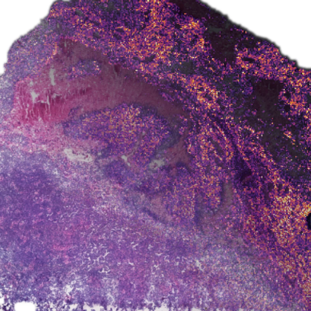
Our lab uses advanced single-cell techniques to understand the biology underlying pediatric brain tumors. Where do they come from during brain development? How do they evolve under pressure from treatment? And how do they develop resistance to the therapies we use?
Learn more about single-cell work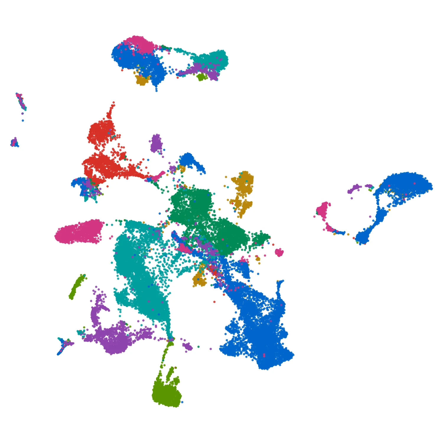
The wiring and rewiring of tumor circuits
Cancer cells rely on complex epigenetic mechanisms to sustain and adapt their core transcriptional circuits. We use cutting-edge chromatin techniques to better understand these relationships and dependencies, and to look for new ways to disrupt them.
Learn more about chromatin and transcription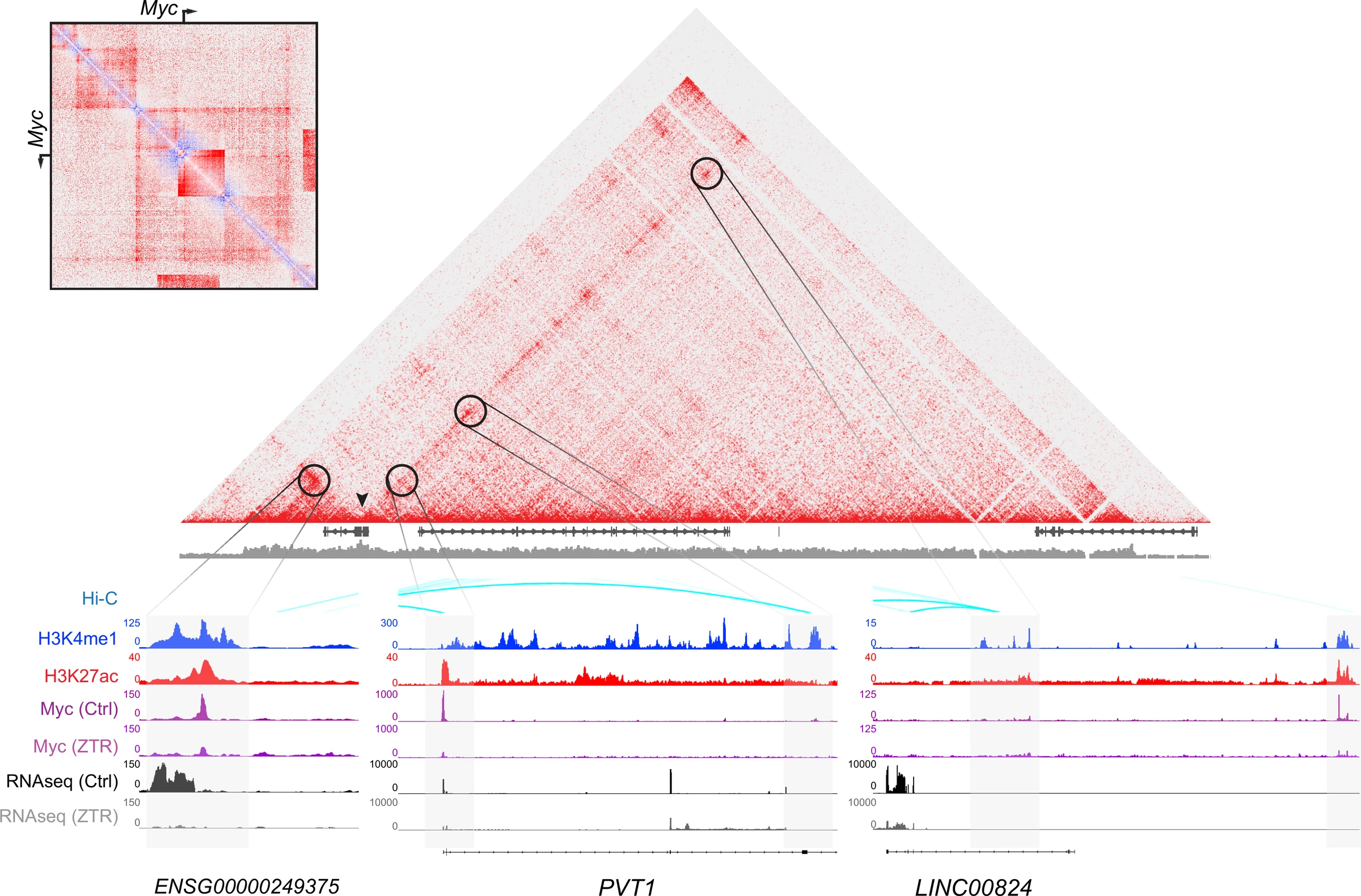
Discovery in a dish
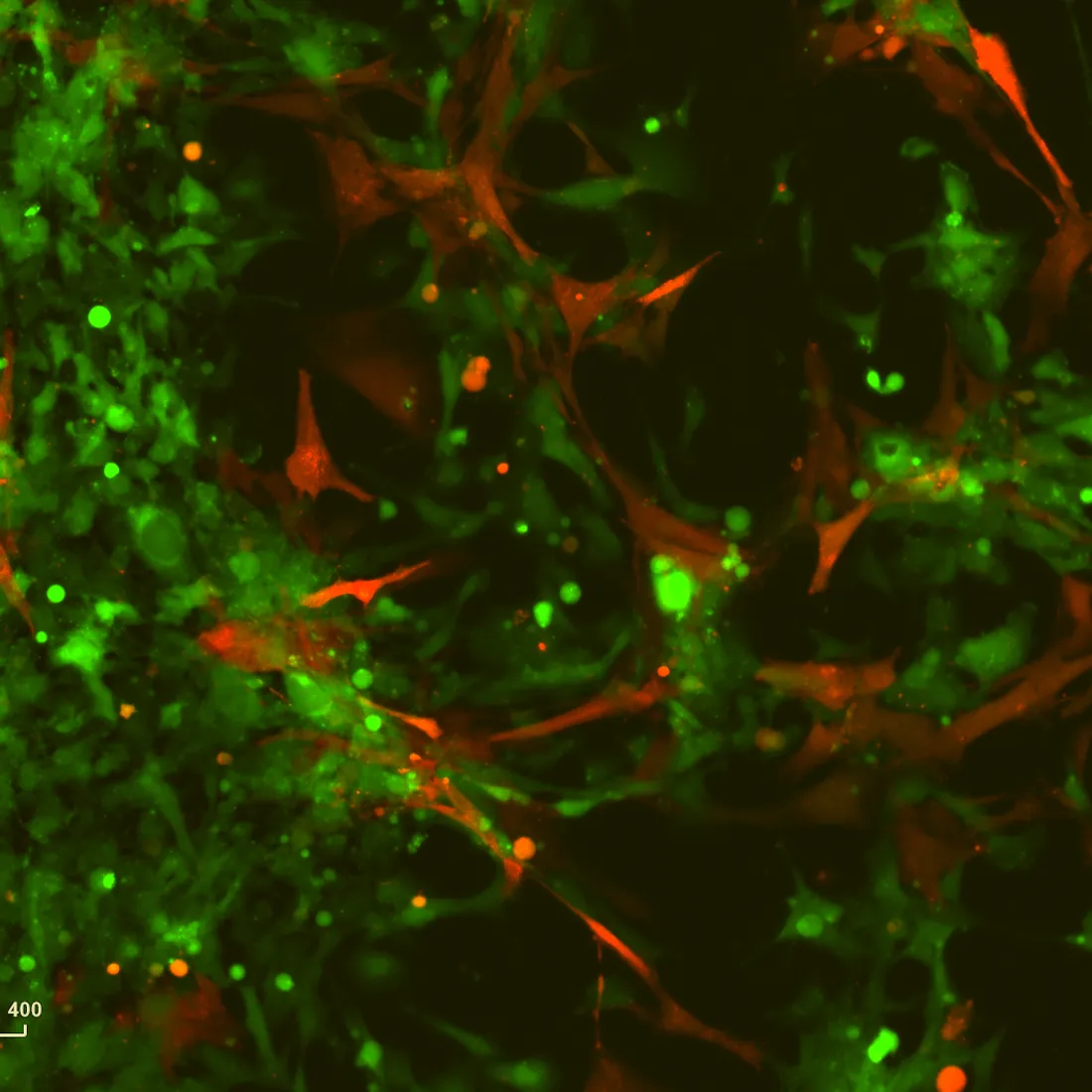
We use both normal and cancer cells to model tumor biology, interactions with the microenvironment, and effects of treatment. How do cancer cells respond to disruptions of their core transcriptional circuits? Do dependencies exist that allow us to effectively kill tumor cells while sparing normal ones?
Learn more about our cell models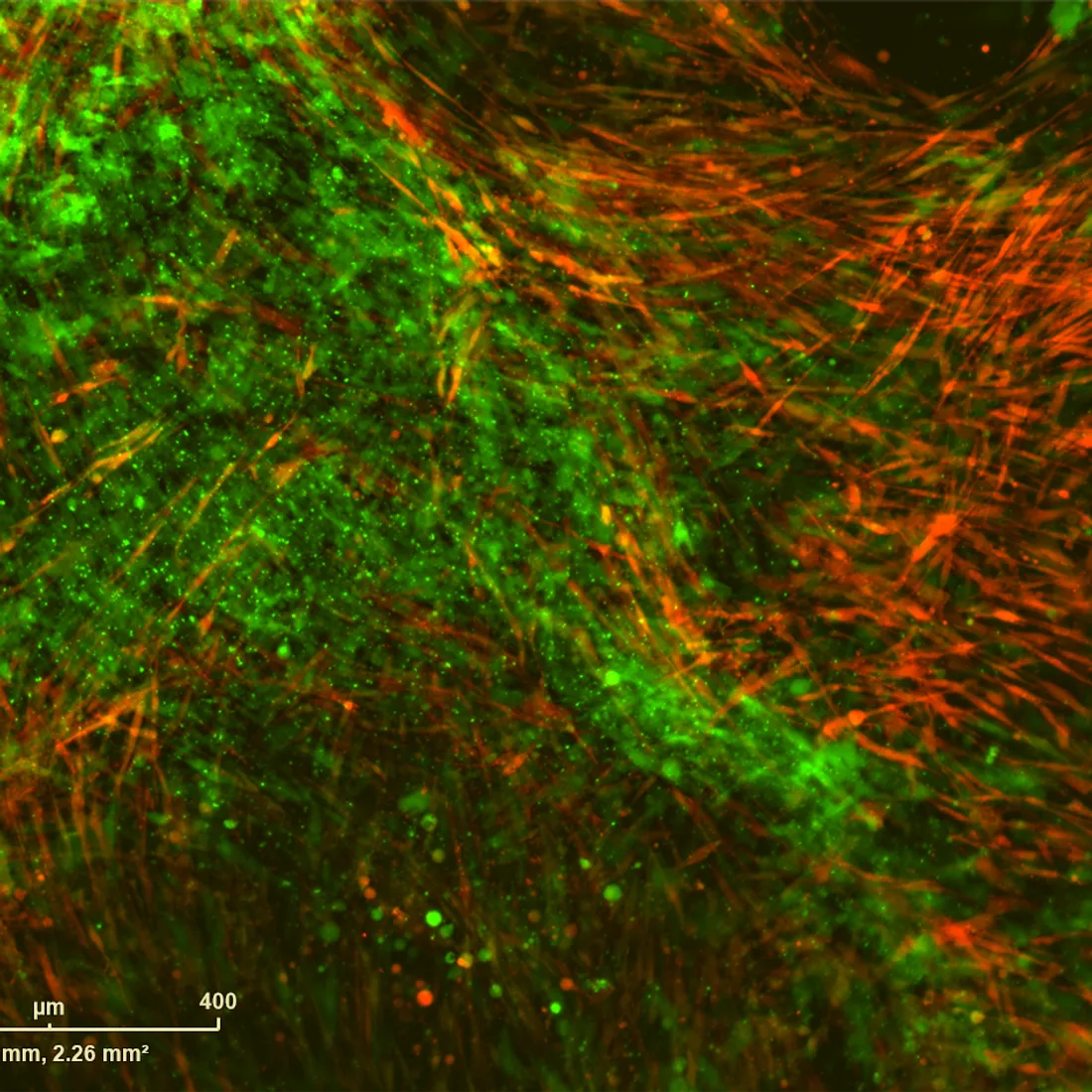
Modeling the next generation of therapy
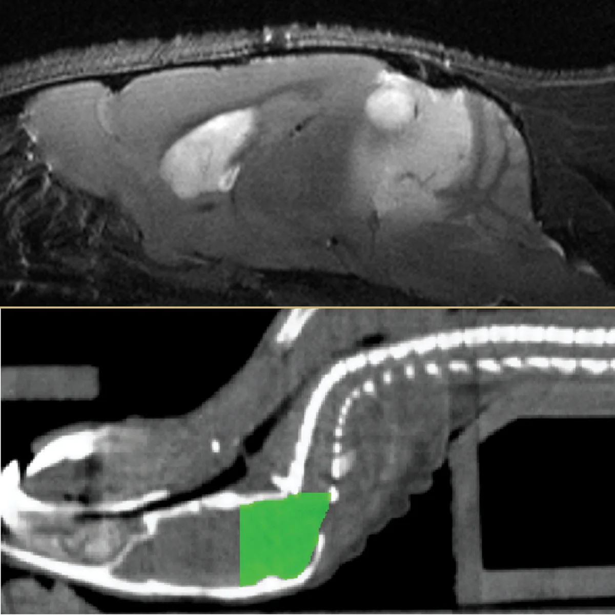
We use sophisticated animal models to test promising cancer treatments. From intracranial drug infusions to CT-guided conformal radiation to volumetric MRIs, we use the most advanced techniques in neuro-oncology to prioritize new agents for clinical trials.
Learn more about preclinical therapy