Research
What we work on
Our lab’s interest across the field of neuro-oncology is broad but anchored around one thread. Can our understanding of fundamental dynamics of chromatin inform unanswered questions in our clinical practice? And most importantly, can we use this framework to nominate new targets for therapy?
Single-cell and spatial biology
Deconstructing tumors, cell by cell
A single tumor is not a single thing. We look at them one cell at a time, looking for insights into where they come from and how they survive the treatments we use.
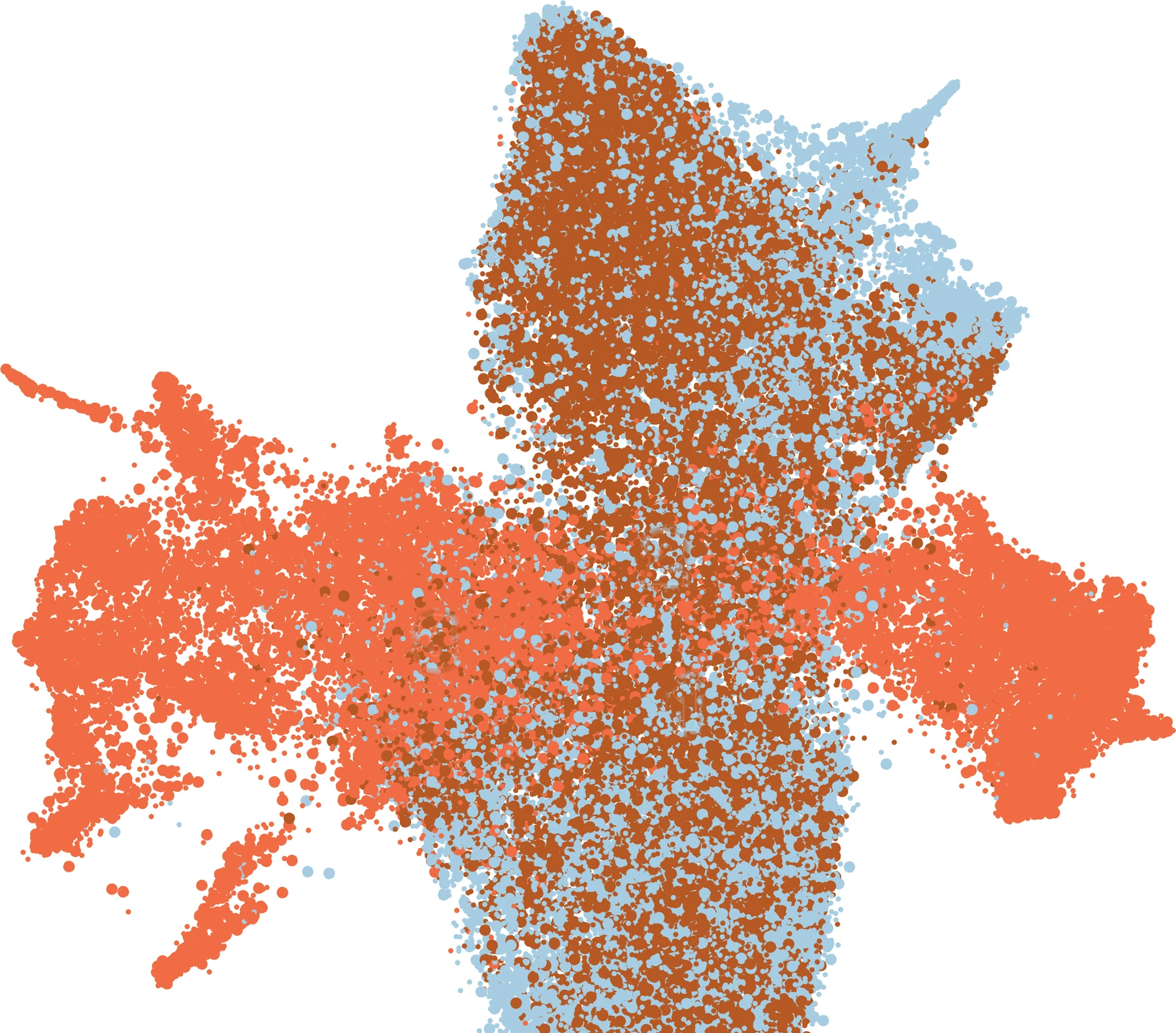
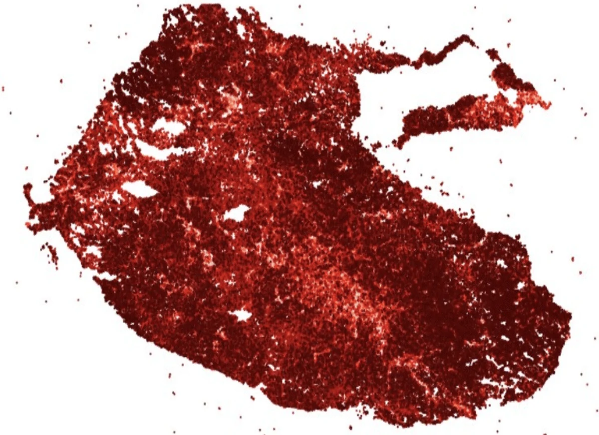
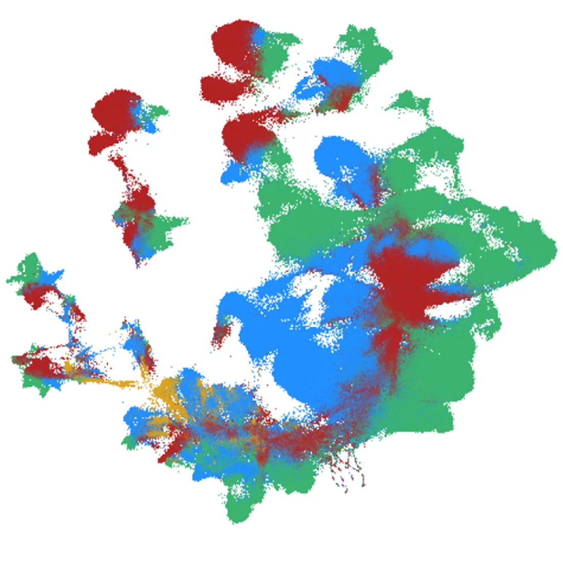
Each individual brain tumor carries a single diagnosis but is comprised of many subclonal populations of cells. These cells harbor distinct identities, fate commitments, and chromatin landscapes. Our lab leverages multiomic chromatin profiling at single cell resolution in order to understand how this more granular view of tumors can inform our understanding of questions relevant to clinical care. We study how features such as enhancers and promoters interact to shape developmental lineages, subclonal heterogeneity, and evolution of resistance under pressure from therapy. The ultimate aim of this avenue of research is to use our understanding of these cellular constraints to better nominate targets for new therapies.
Selected papers
- Single-cell multi-omics identifies metabolism-linked epigenetic reprogramming as a driver of therapy resistance
- Differential phagocytosis induces diverse macrophage activation states in malignant gliomas
- NTRK fusions can co-occur with H3K27M mutations and may define druggable subclones within diffuse midline glioma
Chromatin and transcription
The wiring and rewiring of tumor circuits
Every cell carries the same DNA but reads only part of it. We study how tumors change what gets read — and how they reconfigure once treatment begins.
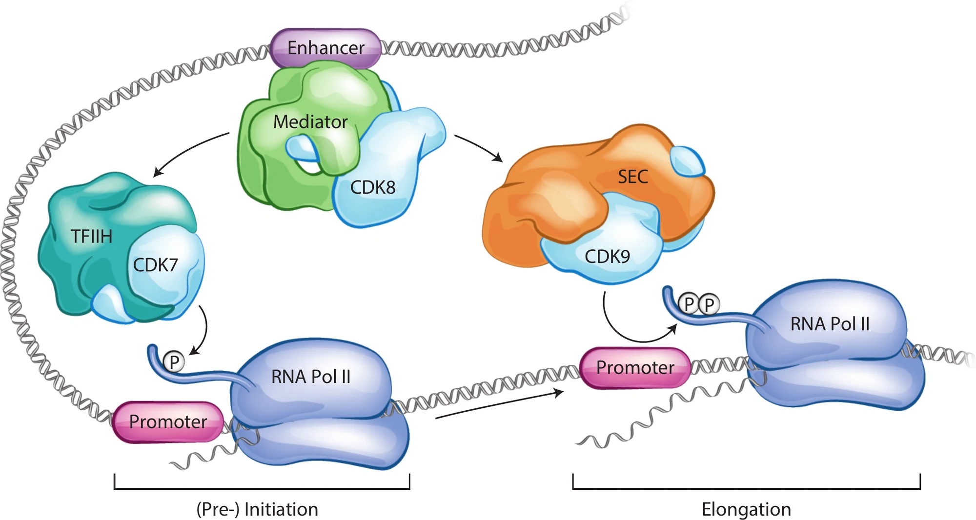
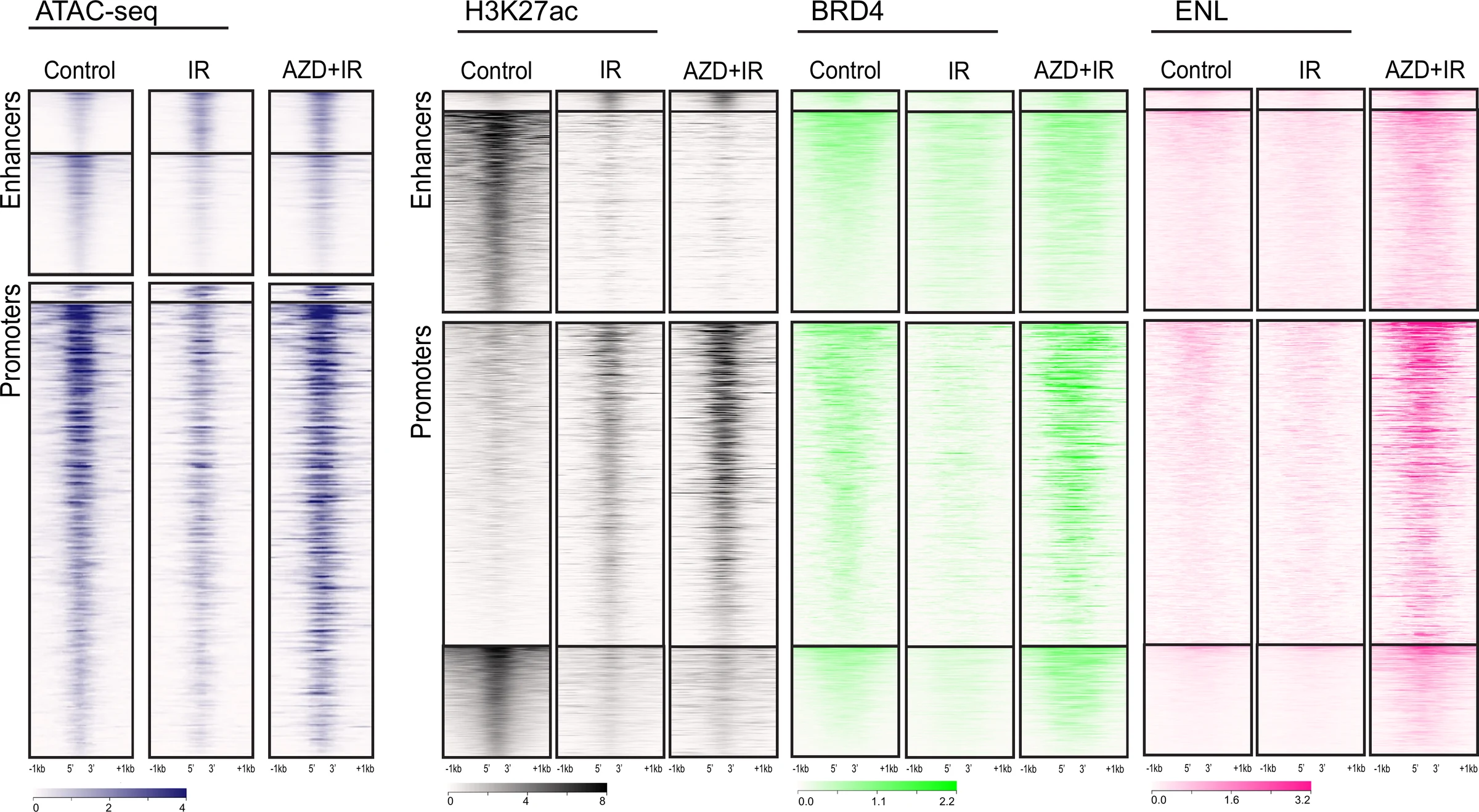
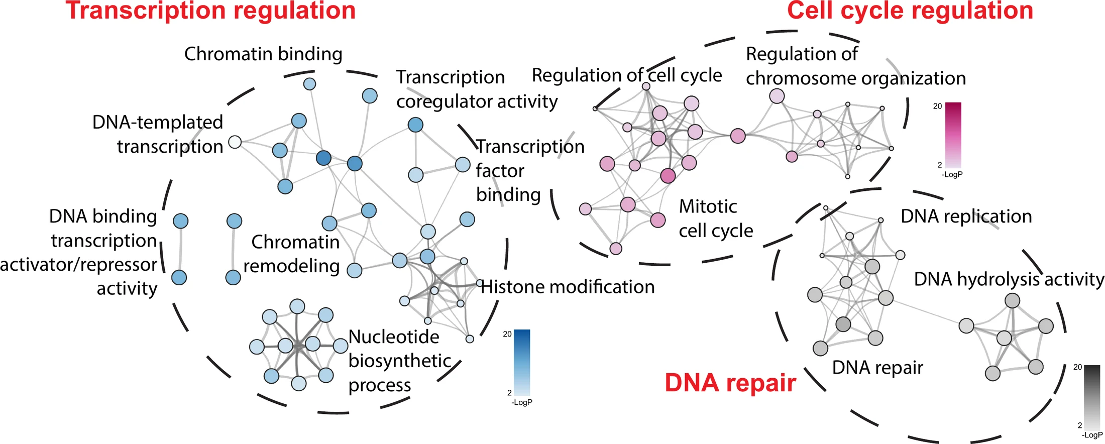
One of the primary areas of focus for our lab is using chromatin frameworks to understand the behavior of tumors. Transcription, or how genes are turned on or off in the appropriate context, is a complex interplay between transcription factors, transcriptional machinery, and epigenetic modifications to chromatin itself. In contrast to adult cancers that are frequently caused by the accumulation of multiple mutations over time, pediatric brain tumors are more often driven by epigenetic alterations. The best example of this may be in H3K27-altered diffuse midline gliomas, where a substitution to a histone tail disrupts epigenetic post-translational modifications genome wide. In other pediatric brain cancers, the core driving alterations converge on transcriptional machinery indirectly. For example, MYC amplifications in high-risk medulloblastomas augment RNA polymerase II activity at the pause-release checkpoint, driving increased transcriptional activity at loci that are primed by cell context to be responsive.
We also use these same techniques to understand how tumors adapt and evolve over time as they undergo treatment. Radiation induces a rapid reorganization of transcriptional machinery, as gliomas turn on the programs required to repair DNA damage and recover from radiation effects. We hope that by better understanding the dynamics underlying these processes, we can identify weaknesses that may be amenable to new kinds of therapeutic interventions.
Selected papers
Model systems and preclinical therapy
Modeling the next generation of therapy
Many drugs that look promising do not work. We try to find that out early, understand why, and identify new compounds that may be more likely to succeed.
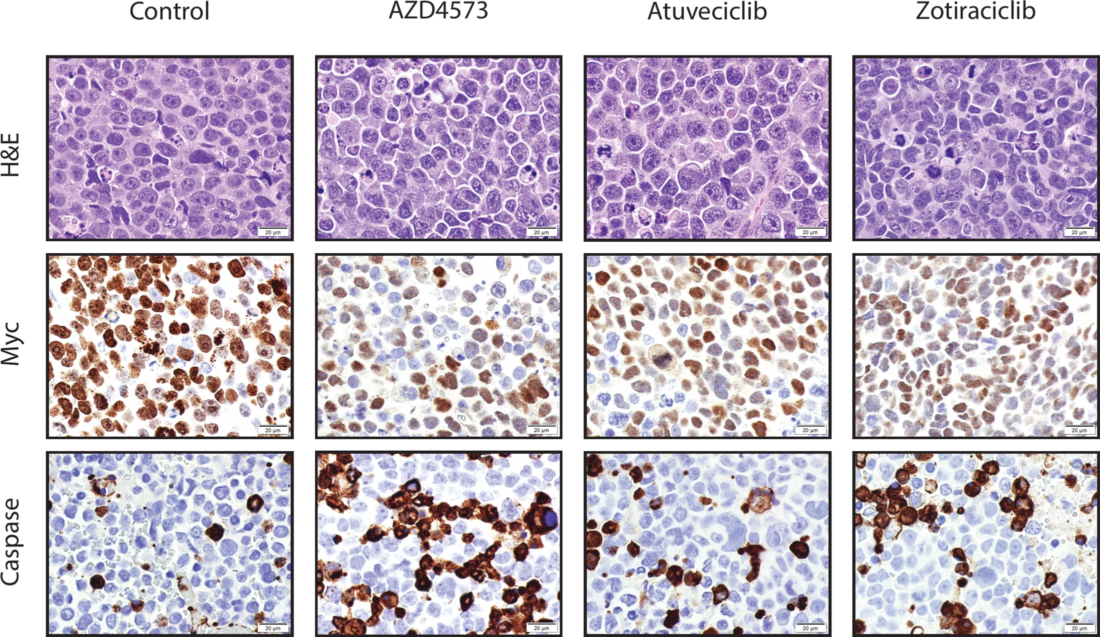
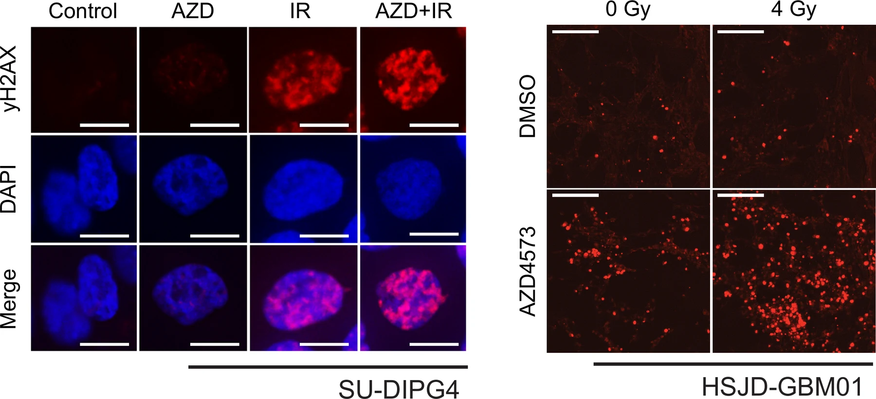
The ultimate aim of our lab is the same as all in this field: to advance better treatments into clinical trials for children with these diseases. Preclinical testing of anticancer therapies is fraught with limitations and to be interpreted with caution. However, we believe that rigorous testing through cell culture, neurosphere, organoid, and animal models can help us ask questions relevant to the patients we care for.
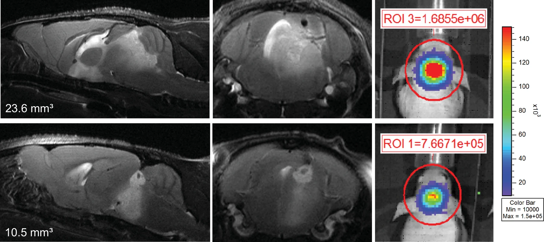
We utilize a range of functional in vitro systems and orthotopic animal models to test new therapies that might benefit children with brain tumors. This allows us to better understand mechanisms of treatments and prioritize drugs with promise of benefit. We test conventional chemotherapies as well as innovative, machine-learning derived compounds, leveraging the expertise across the Morgan Adams Foundation Pediatric Brain Tumor Research Program and the University of Colorado Cancer Center.
Our primary interest has been in transcriptional modulators capable of penetrating across the blood-brain barrier, a major barrier for translational research in neuro-oncology. In parallel, our team has also contributed to the critical evaluation of proteasome inhibitors, metabolic inhibitors, and a range of other targeted small molecule inhibitors or immunotherapies. Ultimately, we aim to nominate and prioritize compounds most likely to provide clinical benefit before advancing to early phase clinical trials.
Selected papers
- Preclinical evaluation of ixazomib for high-risk pediatric brain tumors
- Development of an anti-CD99 antibody enables targeting of diffuse midline glioma
- Venetoclax cooperates with ionizing radiation to attenuate diffuse midline glioma tumor growth
- Super elongation complex as a targetable dependency in diffuse midline glioma
Figures are drawn from published and unpublished work by the lab. See Publications for sources.